
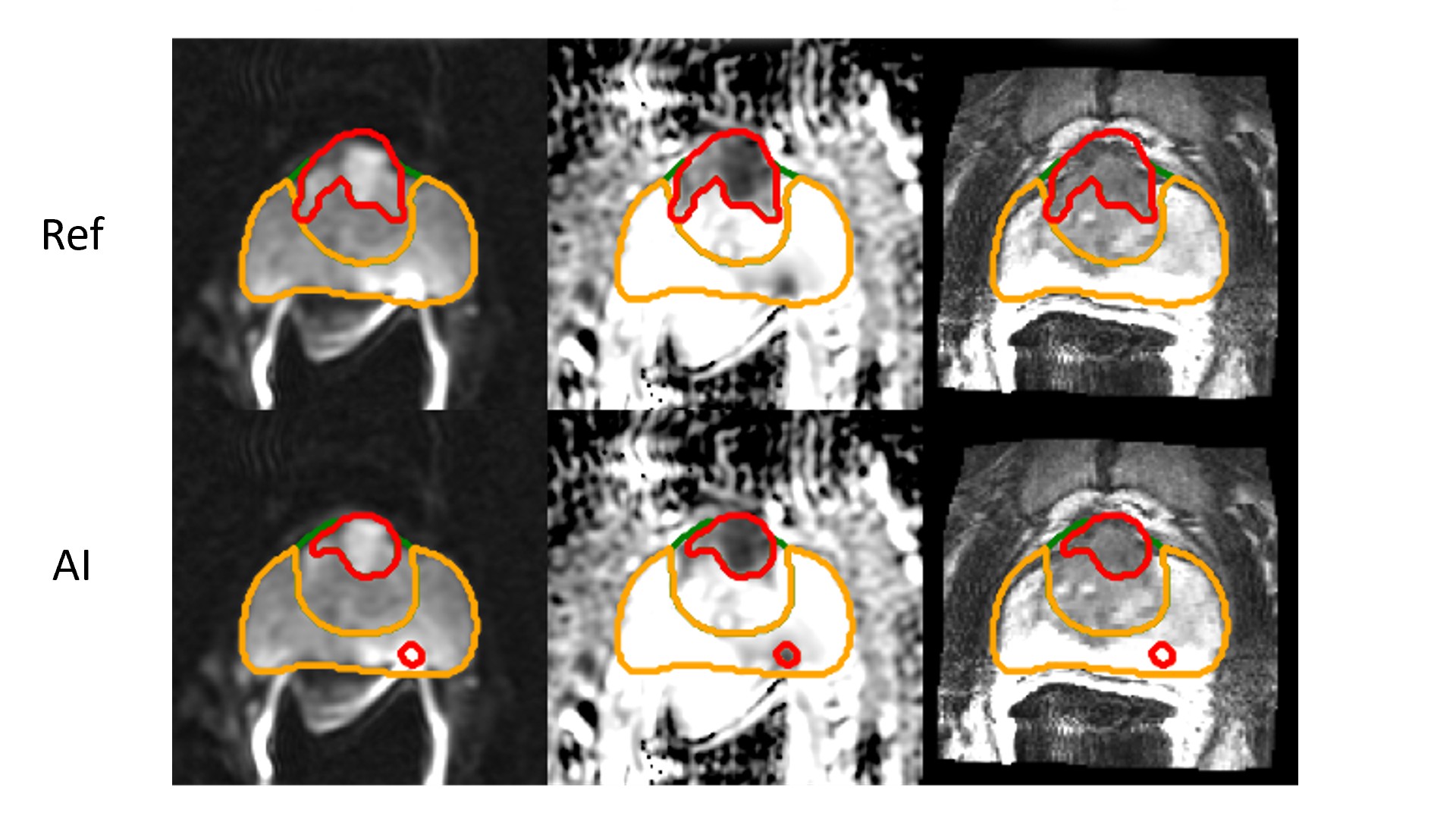
AI-based prostate MRI analysis may help clinicians measure tumor volume faster and use that measurement as one input to risk assessment, but the reported research should not be treated as a replacement for clinical judgment or validated care pathways. A study described by Brigham and Women’s Hospital researchers used image segmentation to outline prostate tumors, calculate their volume, and examine associations between tumor volume and later recurrence or metastasis after surgery or radiation therapy.
What changed in the reported research
The research team developed an AI-driven model to analyze prostate MRI scans with a segmentation approach. The model outlined identified cancer tumors, estimated tumor volume, and evaluated the relationship between those measurements and known patient outcomes over five to ten years.
According to the source record, the researchers trained and tested the model using MRI data from more than 700 cancer patients. They reported that the model identified 85% of aggressive prostate tumors and completed image analysis in seconds. The source also states that its assessment of tumor volume and intraprostatic location was as fast and accurate as human experts. These findings were reported as published in Radiology in October; readers should verify the complete paper, publication date, study design, and evaluation criteria in the journal record.
Why tumor volume matters
The study focused on a practical clinical question: whether existing diagnostic information can provide a clearer view of disease extent and prognosis. The reported findings linked larger tumor volume with a greater likelihood of recurrence or metastasis, including after radiation therapy or surgery. This is relevant because localized and metastatic prostate cancer have materially different outcomes in the survival figures cited by the source.
For care teams, a repeatable tumor-volume estimate could support review of imaging alongside pathology, staging, treatment options, and follow-up planning. For imaging and AI stakeholders, the result illustrates a broader direction in medical AI - moving beyond detection alone toward quantitative measurements intended to inform prognosis.
Decision impact for healthcare AI evaluation
The reported performance is not sufficient on its own to justify deployment. Organizations considering a similar workflow should distinguish between a research model, a locally validated clinical tool, and a product authorized for a specific clinical use. The supplied record does not establish regulatory status, commercial availability, interoperability, clinical approval, or performance across sites and MRI protocols.
- Confirm the model’s intended use, patient population, MRI acquisition requirements, and reference standard in dated official documentation.
- Review the full study for cohort composition, definitions of aggressive tumors, outcome measures, validation design, and confidence intervals.
- Run a local evaluation against representative MRI studies, including scanner, protocol, image-quality, and demographic variation relevant to the intended setting.
- Define radiologist and urologist review responsibilities, escalation rules, audit trails, and how volume estimates will be presented without overstating prognostic certainty.
Technical context and implementation limits
The research workflow reportedly used PyTorch on an NVIDIA GeForce RTX 3070 GPU and the open-source nnUNet algorithm for image segmentation. This identifies the research environment, not a required or sufficient production architecture. Hardware selection, inference latency, cybersecurity controls, data governance, model versioning, and integration with imaging systems must be assessed separately for any implementation.
The source itself notes that the number of MRI scans examined was relatively limited and characterizes the findings as early but encouraging. Associations between AI-estimated volume and later outcomes do not establish that the model independently determines prognosis, nor do they demonstrate benefit for every patient or treatment pathway. Prospective validation and project-specific testing remain necessary.
FAQ
Did the reported AI model diagnose prostate cancer by itself?
No. The source describes a model that analyzes prostate MRI, segments identified tumors, and estimates tumor volume. It does not establish that the model should independently diagnose cancer or make treatment decisions.
Can a hospital reproduce the reported results using nnUNet and an RTX 3070 GPU?
Not necessarily. The source identifies nnUNet, PyTorch, and an NVIDIA GeForce RTX 3070 GPU as components of the study setup, but it does not provide the complete dataset, preprocessing method, training configuration, validation protocol, or deployment requirements. Those details must be verified through the full research publication and local testing.
Conclusion
The reported study suggests that AI segmentation of prostate MRI can rapidly quantify tumor volume and may contribute to recurrence or metastasis risk assessment. Its main decision value is as evidence for further clinical and technical evaluation, not as proof that an unverified model is ready for routine use.
After reviewing AI MRI Analysis for Prostate Cancer Risk Assessment, continue with related solutions for related evaluation paths.

 WeChat
 Profile