
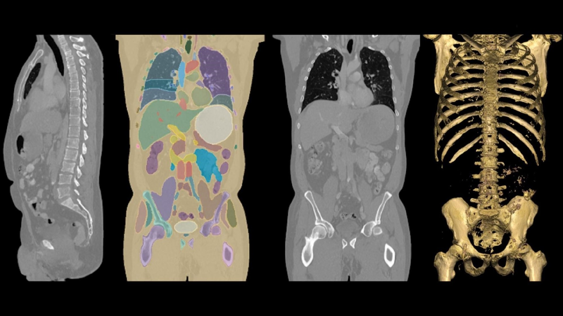
MONAI Core v1.4 expands the MONAI medical AI framework with VISTA-3D, VISTA-2D and MAISI, while VISTA-3D and MAISI are also offered as NVIDIA NIM microservices. For teams moving from medical imaging research toward operational workflows, the release presents both open-source model access and a containerized inference path. Model fit, local data performance, workflow integration and applicable clinical requirements still need to be evaluated for each project.
What MONAI Core v1.4 adds
MONAI Core v1.4 introduces additional algorithm capabilities and three foundation models: VISTA-3D, VISTA-2D and MAISI. MONAI originated as a collaboration between NVIDIA and King’s College London and has developed into an ecosystem for medical imaging AI research. The supplied announcement reports more than 3.5 million downloads and more than 1,000 published papers over MONAI’s first five years.
The practical value of the v1.4 release is that it addresses several distinct imaging tasks within one framework:
- VISTA-3D targets interactive anatomical annotation and segmentation in 3D CT images.
- VISTA-2D supports microscopy analysis for cell biology research.
- MAISI is designed for synthetic 3D CT image generation.
These models should not be treated as interchangeable. A CT segmentation workflow, microscopy workflow and synthetic-data workflow have different data inputs, evaluation criteria, human-review requirements and integration risks.
Capabilities by model
VISTA-3D is described as an interactive foundation model for annotating anatomy in 3D CT images. The announcement states that it covers more than 126 anatomical classes out of the box and can use interactive annotations to segment previously unseen structures in a zero-shot setting. Teams assessing VISTA-3D should test the relevant anatomy, CT acquisition characteristics and user interaction process rather than assuming that reported coverage matches their intended use.
VISTA-2D uses a Transformer architecture of approximately 100 million parameters built on Meta’s Segment Anything Model architecture. It is intended to process multiple cell types and imaging modalities. The source cites validation on TissueNet, LIVECell and Cellpose datasets; however, those datasets do not establish performance for a specific laboratory’s instruments, staining protocols or image quality.
MAISI is a latent diffusion model for synthetic medical imaging. According to the announcement, it can generate 3D CT images at resolutions up to 512 × 512 × 768 voxels, with voxel sizes from 0.5 mm to 5.0 mm. Users can control 10 body-habitus classes and corresponding segmentation masks. Before using synthetic images for development or evaluation, organizations should establish whether the generated data preserves the characteristics needed for their intended task and whether its use is appropriate under their data-governance process.
NIM deployment path for VISTA-3D and MAISI
VISTA-3D and MAISI are available as NVIDIA NIM microservices within NVIDIA AI Enterprise. The announcement describes these as containerized, GPU-accelerated inference services that can be deployed across cloud, data center and workstation environments. It also identifies TensorRT-optimized inference engines, industry-standard APIs, GPU-system optimization for latency and throughput, flexible infrastructure deployment, and enterprise security and scalability features.
For an implementation evaluation, begin with a defined imaging use case and an approved data path. Then confirm the exact model version, NIM container requirements, GPU compatibility, API behavior, authentication model, observability needs and workflow interfaces in dated NVIDIA documentation and the complete deployment bill of materials. A pilot should measure task-specific quality, turnaround time, infrastructure utilization and failure handling under representative local conditions.
Where the release may fit
Research teams may use MONAI Model Zoo access and the open framework to investigate segmentation, microscopy analysis or synthetic imaging. Organizations seeking an operational inference packaging option may assess the VISTA-3D and MAISI NIM microservices alongside their existing medical imaging systems. The source also describes M3 and VILA-M3 as a research initiative that combines visual understanding with natural-language processing, as well as Holoscan SDK integration for radiology workflows, real-time surgical guidance and surgical robotics contexts.
The announcement does not establish diagnostic accuracy, regulatory clearance, clinical safety, interoperability with a particular PACS or EHR, or suitability for autonomous clinical decisions. Those matters require review of official documentation, local governance requirements and project-specific validation.
FAQ
Is VISTA-3D limited to fully automatic CT segmentation?
No. The source describes VISTA-3D as an interactive model for 3D CT annotation. Its stated zero-shot capability uses interactive annotations to learn segmentation of new structures. The required level of user input and resulting quality should be tested on the intended workflow.
Can MAISI-generated CT images replace real clinical data?
The source describes MAISI as a model for creating synthetic 3D CT images, not as a replacement for real clinical data. Teams should define the permitted purpose, assess generated outputs against relevant data characteristics and validate any downstream model or workflow with appropriate real-world data.
Conclusion
MONAI Core v1.4 provides a broader medical AI toolkit spanning 3D CT segmentation, 2D microscopy analysis and synthetic CT imaging. VISTA-3D and MAISI NIM microservices add a containerized deployment option, but adoption decisions should be based on documented configuration requirements and evidence from a controlled, use-case-specific evaluation.
After reviewing MONAI Core v1.4, VISTA-3D and MAISI for Medical AI Workflows, continue with buyer selection questions for related evaluation paths.

 WeChat
 Profile